Pioneering surgery schoolgirl loses her legs and learns to skateboard.
I am so grateful to Mr Marks and his team for the amazing work they did to save Rosie's life. It is just wonderful to see her see her living life to the full.
idiopathic

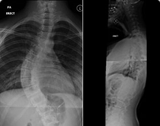
This is the commonest type of scoliosis. ‘Idiopathic’ means that there no known cause. This is usually a diagnosis of exclusion, based on a normal MRI scan of the full spine. A genetic cause is currently the focus of research as the underlying cause of this type of scoliosis.
Based on the age of onset it is classified as Infantile (onset between birth and 3 years); Juvenile (3 – 7 years); Adolescent (7 – 18 years). More recently we classify these as Early-onset (under 5 years) and Late onset (over 5 years).
Early-onset scoliosis has several distinctive features. This type of scoliosis is commoner in boys, are more commonly left sided thoracic curves. It is less common in girls. A right-sided curve in a girl is more likely to progress.
90% of this type of scoliosis corrects itself with growth (non-progressive). The 10% that are likely to progress can sometimes be predicted based on the radiographic features (RVAD: rib-vertebral angle difference, also known as the Mehta angle). Though more often the progression is detected by performing serial radiographic assessments.
Late-onset / Adolescent scoliosis: This type of scoliosis is common in teen-aged girls. There can be one or more curves. The main curve is known as the structural curve and the other flexible curves above and below it are called compensatory curves. There can be more than one structural curve. These are usually determined on xrays. Progression of the scoliosis is closely linked to the age at presentation and residual skeletal growth. The onset of menarche is a marker of the adolescent growth spurt. Growth spurt is assessed by the onset of periods and by the Risser sign on pelvic xrays. (Risser pic) Close monitoring for the progression, especially during the growth spurt, aids in planning treatment. Genetic cause is currently under investigation. It is not unusual for a family member or a sibling to have this condition.
Curves under 30 degrees are observed and monitored. Treatment of the curves between 30 and 50 degrees is subject to discussions with the patients and family and may be treated with a brace to prevent progression. Curves over 50 degrees may require surgery. This may be performed either anterior, posteriorly or a combination, based on various factors.